Corneal topography and pachymetry
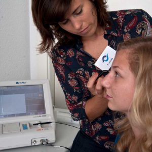
A-SCAN (MEASUREMENT OF THE AXIAL LENGTH
A-scan biometry is a measuring device for measuring the length of the eye using ultrasound.
After applying an anesthetic drop to the cornea, a small pen (ultrasound probe) touches the cornea and measures the eye length.
PACHYMETRY
A pachymeter is a device used for the exact measurement of corneal thickness (accuracy 1 to 5 micrometers (μm) This is relevant for the correct determination of the intraocular pressure by tonometry, since the measured value is influenced by the corneal thickness in most common methods. With the result of the corneal thickness the actual intraocular pressure can be calculated. The standard value of a cornea is approximately 550 μm.
After applying an anesthetic drop to the cornea, a small pencil touches the corneal surface and measures the thickness using ultrasound.
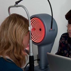
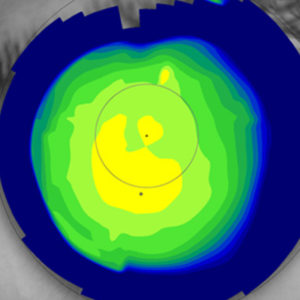
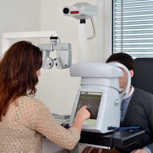
CORNEAL TOPOGRAPHY (SIRIUS)
This examination serves to measure the front and back surface of the cornea. It can be used to determine, for example, astigmatism or a thinning of the cornea with a bulge (keratoconus). This procedure is also used for contact lens fitting which guarantees an optimal fit of the lens. In cases of severe astigmatism, special lenses can be used during cataract surgery as a result of the topography.
The corneal surface is individual for each person. The anterior and posterior surface of the cornea is measured topographically, so that a relief of the cornea can be calculated. This topography is displayed graphically.
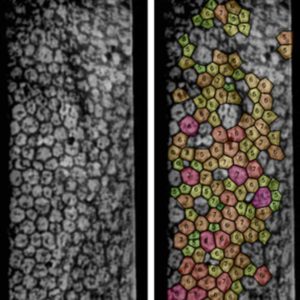
ENDOTHELIAL CAMERA (PERSEUS)
If the number of cells decreases or the cell size changes, there is a risk that the cornea becomes cloudy.